
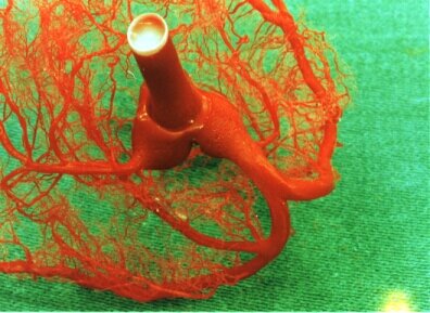
Cast of the coronary system of a pig heart. From the aorta casting material was infused retrograde into the coronary arteries. The aortic valve prevented flux into left ventricle. Tissue was then dissolved by acid.
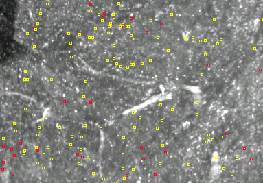
Image of part of a cutting plane of a goat heart under normal light. Vessels are filled with paint and some vascular structures are clear. The red and yellow squares identify the positions of red and yellow fluorescent microspheres
In case coronary arteries get partially obstructed by arterioscleroses the resistance vessels relax to compensate for the pressure drop induced by the obstruction. This may bring areas within the heart muscle into problems by having a supply of blood that is not sufficient for sustaining muscle cells at an active state or even alive.
This project aims to relate distribution of blood flow to the structure of the coronary arterial tree and has experimental as well as theoretical aspects. For the experiments an imaging cryo-microtome is used.
Under relevant experimental conditions fluorescent microspheres are injected into the coronary artery which distribute with flowing blood. After the experiment the coronary vessels are filled with a fluorescent dye. The heart is then frozen and mounted in the cryo-microtome and sliced. After each slice digital images are taken with appropriate lighting and filters of the cutting plane. In this way a reconstruction of the arterial tree can be made as well the position of the microspheres automatic detected.
The theoretical aspect is related to understand the distribution of these microspheres in relation to the structure of the vascular bed. Hence, the reconstruction forms the basis of predicting flow distribution in a variety of conditions relevant to health and disease. The reconstructed tree allows also the evaluation of control properties measured in isolated vessels, an other project in the department of Medical Physics. Quit a bit of this theoretical work has already been done on trees that were generated from certain design principles. However, these studies show that a more realistic representation of the tree is needed.

the Netherlands Heart Foundation
![]()