
|
 |
| Lines of research |
|
| [ Medical Imaging ] [ Minimal Invasive Surgery ] |
The research in the field of Medical Imaging is focused on the physics of image formation, image analysis and visualization. The research activities are carried out in close cooperation with clinical partners. Projects focus on the following themes: |
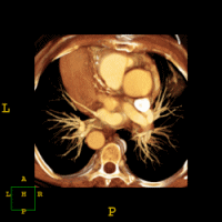 |
With spiral CT a large volume of image data can be acquired within a short period of time. This has made CT angiography feasible, a technique in which blood vessels are visualized after injection of a contrast medium. Matching techniques are investigated to register data sets acquired before and after contrast injection, for example of the renal and cerebral arteries, in order to improve the visualization of these arteries and to aid the detection of vascular pathology. Research also takes place into the possibilities and limitations of CT with a very low radiation dose.
Recently, an experimental imaging technique for the determination of viscoelastic properties of tissue has been introduced: MR Elastography. The viscoelastic characterization of tissue is expected to be a useful indicator for malignant tissue. The tissue properties are extracted from image data obtained via a MR sequence that is coupled to a mechanical harmonic excitation of the tissue. Research on MR Elastography is focused on development of instrumentation for the excitation of tissue and image analysis methods for extraction of the viscoelastic properties from the image data. |
|
In reconstructive orthopedic surgery of joint structures there is a need for preoperative prediction of intra-operative joint geometry and evaluation of postoperative joint function. To obtain the necessary information for evaluation and prediction we combine 3D imaging techniques, dedicated 3D image analysis methodology, biomechanical modeling of joints and force-motion measurements of joints. The imaging part of this project addresses the development of methods for obtaining the geometry and the mechanical properties of the anatomical structures within the joint.
|
Contact
Dr. H.W. Venema
Dr. ir. G.J.Streekstra
Prof. dr. ir. C.A. Grimbergen
Key Publications
|
In close co-operation with the Cardiovascular Physics group within our department and the Netherlands Institute for Brain Research we develop methods to detect topological and geometrical properties of coronary vessel trees. For this purpose 3D confocal microscope images of the arteriolar bed are obtained. For measurement of topology and detection of the length and diameter of vessel segments from the image data procedures are under development. The combination of topological and geometrical data will be useful for modeling flow distributions in the coronary vasculature. |
|

|
| MAIN PAGE MEDICAL TECHNOLOGY |
|