
Applications of Image Registration Techniques in Spiral Computed
Tomography (CT)
With multislice spiral CT (Fig. 1) it is possible to scan a large volume
of the patient within a short amount of time and with a high spatial
resolution in all directions. One of the applications of multislice
spiral CT is CT angiography (CTA). With CTA various arteries in the
patient can be made visible in a minimally invasive way. To fully
comprehend the complex geometry of the vascular structure, the CTA data
sets are often visualized by making a maximum intensity projection (MIP)
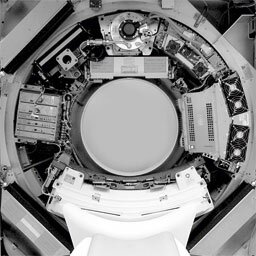
Figure1: View of a multislice CT scanner with the front cover removed.
as shown in Figs. 2-4. In a CTA data set, however, the maximum value in
the direction of projection is often that of bone or arterial wall
calcifications. Therefore the voxels that represent bone and obscure the
vascular structures of interest have to be removed from the data set,
prior to the generation of the MIP.

Figure 2: Coronal MIP image of the aorta and the renal arteries. The white
structures are arterial wall calcifications.
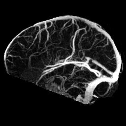
Figure 3: Sagittal MIP image of the cerebral veins.
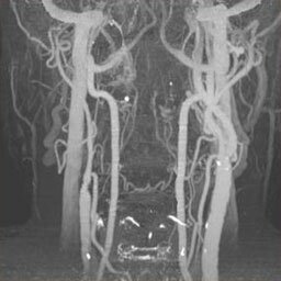
Figure 4: Coronal MIP image of the carotid arteries.
The removal of bone should be performed in such a way that the
arteries are not affected. We therefore introduce the use of an
additional scan which is made
prior to the injection of contrast agent, in which the bone can be
identified unambiguously. The bone is removed by determining the
bone voxels in the (registered) nonenhanced data set and giving
the corresponding voxels in the contrast-enhanced data set an
arbitrary low value. This method, called Matched Mask Bone
Elimination (MMBE), has been introduced by Venema et. al.
(Radiology 2001; 218:893-898) for CTA examinations of the Circle
of Willis.
Currently the application of the MMBE method is
expanded to the neck region. The rigid registration method that
was used for the region of the Circle of Willis no longer
suffices, because the head and neck region contains bone
structures that can move with respect to each other. A watershed
algorithm to separate the bones followed by a piecewise rigid
registration procedure are introduced to solve this problem (Fig. 5).
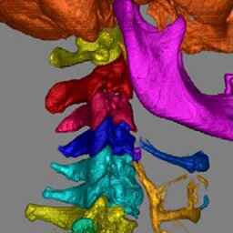
Figure 5: Volume rendering of the output of the watershed algorithm. Each identified bone is individually marked with an unique color.
The image quality is investigated as a function of scan and
reconstruction parameters with the aid of a number of phantom studies
(Fig. 6). These phantom studies allow us to optimize the scan protocol
when scanning patients and to understand the physical and mathematical
properties of a CT scanner and its reconstruction software.
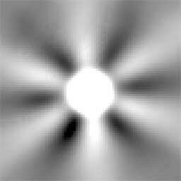
Figure 6: Multislice spiral CT scan of a small, dense sphere. The artifacts clearly present in this image are also of importance in clinical scans.

Marcel van Straten was born on December 21st, 1974 in Rotterdam, The
Netherlands. In 1993 he started his study Applied Physics at the Faculty
of Applied Sciences at the Delft University of Technology. His final
project, performed at the Magnetic Resonance Imaging group, was entitled
'Simulation and in vitro measurement of relaxation effects of
paramagnetic contrast agents'. In September 1999 he received the degree
of Master of Science in both the main variant and the business variant.
In December 1999 he started his Ph.D. project at the Department of
Medical Physics and at the Department of Radiology of the University of
Amsterdam in the Academic Medical Center under the supervision of Prof.
dr ir C.A. Grimbergen, Prof. dr G.J. den Heeten and dr H.W. Venema. The
main focus of this project is the application of image registration
techniques in spiral CT. Philips financially supports this project since
December 2003.
![]()